
Osteoblast precursors and inflammatory cells arrive simultaneously to sites of a trabecular-bone injury
DOI:
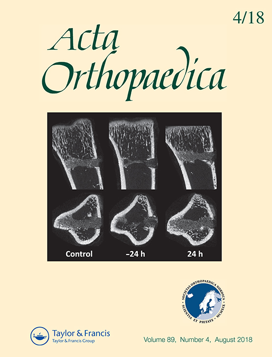
https://doi.org/10.1080/17453674.2018.1481682Abstract
Background and purpose — Fracture healing in the shaft is usually described as a sequence of events, starting with inflammation, which triggers mesenchymal tissue formation in successive steps. Most clinical fractures engage cancellous bone. We here describe fracture healing in cancellous bone, focusing on the timing of inflammatory and mesenchymal cell type appearance at the site of injury Material and methods — Rats received a proximal tibial drill hole. A subgroup received clodronate-containing liposomes before or after surgery. The tibiae were analyzed with micro-CT and immunohistochemistry 1 to 7 days after injury. Results — Granulocytes (myeloperoxidase) appeared in moderate numbers within the hole at day 1 and then gradually disappeared. Macrophage expression (CD68) was seen on day 1, increased until day 3, and then decreased. Mesenchymal cells (vimentin) had already accumulated in the periphery of the hole on day 1. Mesenchymal cells dominated in the entire lesion on day 3, now producing extracellular matrix. A modest number of preosteoblasts (RUNX2) were seen on day 1 and peaked on day 4. Osteoid was seen on day 4 in the traumatized region, with a distinct border to the uninjured surrounding marrow. Clodronate liposomes given before the injury reduced the volume of bone formation at day 7, but no reduction in macrophage numbers could be detected. Interpretation — The typical sequence of events in shaft fractures was not seen. Mesenchymal cells appeared simultaneously with granulocyte and macrophage arrival. Clodronate liposomes, known to reduce macrophage numbers, seemed to be associated with the delineation of the volume of tissue to be replaced by bone. Most fracture healing studies in animal models concern cortical bone in shafts. However, most fractures in patients occur in cancellous bone in the metaphysis, such as the distal radius or in the vertebrae. A growing body of evidence suggests that there are important differences between the healing processes in cortical and cancellous bone.Downloads
Download data is not yet available.
Downloads
Additional Files
Published
2018-07-04
How to Cite
Bernhardsson, M., & Aspenberg, P. (2018). Osteoblast precursors and inflammatory cells arrive simultaneously to sites of a trabecular-bone injury. Acta Orthopaedica, 89(4), 457–461. https://doi.org/10.1080/17453674.2018.1481682
Issue
Section
Articles
License
Copyright (c) 2018 Magnus Bernhardsson, Per Aspenberg

This work is licensed under a Creative Commons Attribution 4.0 International License.
Acta Orthopaedica (Scandinavica) content is available freely online as from volume 1, 1930. The journal owner owns the copyright for all material published until volume 80, 2009. As of June 2009, the journal has however been published fully Open Access, meaning the authors retain copyright to their work. As of June 2009, articles have been published under CC-BY-NC or CC-BY licenses, unless otherwise specified.


PlumX (by Elsevier) is an altmetrics platform that tracks and visualizes the online attention, usage, captures, citations, and social media engagement.

