Time of diagnosis of congenital upper limb anomalies: a cohort study of 703 patients from a local registry in Norway
Ida N SLETTEN 1, Mona I WINGE 1, and Jarkko JOKIHAARA 2
1 Division of Orthopaedic Surgery, Oslo University Hospital, Norway; 2 Department of Hand Surgery, Tampere University Hospital and Tampere University, Finland
ORCIDs, if available, can be found on the article page (https://actaorthop.org/actao/article/view/46807)
Background and purpose — Several European countries offer nationwide prenatal ultrasound screening for fetal anomalies. Few studies have examined prenatal detection of upper limb anomalies, and existing reports originate exclusively from high‑volume centers outside Europe. We aimed to assess the timing of diagnosis in patients with all Oberg–Manske–Tonkin diagnoses except tumorous dysplasias referred to the largest specialized unit in Norway, investigate prenatal detection rates, and explore associations between patient‑ and hospital‑related factors and prenatal detection.
Methods — We extracted data on timing of diagnosis according to Oberg–Manske–Tonkin phenotype, and patient- and hospital-related variables from the CULA (congenital upper limb anomaly) North Oslo Registry from 2018 to 2025. For patients with anomalies detected prenatally or at birth, we analyzed associations between patient- and hospital-related factors and prenatal detection rate by multivariable logistic regression.
Results — 703 consecutive patients were enrolled. 402 (57%) patients had their anomaly detected at birth and 252 (36%) later in life. Prenatal ultrasonography detected the upper limb anomaly in 49 (7.0%) of the patients. Among 426 patients with anomalies visible at birth whose mothers had at least 1 prenatal ultrasound scan, the examination detected the anomaly in 49 (12%). Among phenotypes observed in more than 5 patients, congenital contractures (amyoplasia, distal arthrogryposis), reduction deficiencies (transverse, radial, ulnar), and ulnar polydactyly had the highest detection rates. Prenatally detected cases were more often born in university hospitals and more frequently had bilateral upper limb involvement and/or associated anomalies than those diagnosed at birth.
Conclusion — 7.0% of the upper limb anomalies were detected prenatally, 57% at birth, and 36% later in life. Prenatal detection rates were lower than those reported from larger non-European hand units, likely due to the inclusion of all upper limb anomaly diagnoses in this study and Norway’s decentralized prenatal care system.
Citation: Acta Orthopaedica 2026; 97: 678–684. DOI: https://doi.org/10.2340/17453674.2026.46807.
Copyright: © 2026 The Author(s). Published by MJS Publishing – Medical Journals Sweden, on behalf of the Nordic Orthopedic Federation. This is an Open Access article distributed under the terms of the Creative Commons Attribution 4.0 International License (https://creativecommons.org/licenses/by/4.0/)
Submitted: 2026-06-06. Accepted: 2026-08-23. Published: 2026-09-17.
Correspondence: ida.sletten@icloud.com
All authors designed the study protocol. MIW and INS collected data. INS analyzed data, and all authors interpreted the findings. INS wrote the first draft of the manuscript, and all authors participated in manuscript editing.
The authors would like to thank research coordinators Elise Berg Vesterhus and Silje Stuksrud from the Division of Orthopaedic Surgery at Oslo University Hospital for their administrative assistance.
Handling co-editors: Ilkka Helenius and Robin Christensen.
Acta thanks Eeva Astrid Koskimies-Virta and other anonymous reviewers for help with peer review of this manuscript.
Congenital upper limb anomalies (CULA) are rare [1]. They may be obvious at birth (e.g., polydactyly, syndactyly, severe limb reduction deficiencies) or become evident later in life (e.g., camptodactyly, Madelung deformity). Several CULAs can be detected at prenatal ultrasound [2-6]. The optimal window for visualizing the fetal hand and digits is the late first and early second trimesters, when the hands are sufficiently large, the digits are extended, and intrauterine space allows adequate imaging [2].
Two centers, in Australia and the USA, have reported prenatal CULA detection rates, ranging from 21% to 42% [4-6]. Higher detection rates have been associated with a positive family history, anomalies affecting the entire upper extremity, the presence of associated anomalies, and examinations performed in high-volume centers [3,4,6]. Norway, in contrast, is a sparsely populated country with few high-volume obstetric centers. No prior Norwegian data exists on prenatal CULA detection, and, to our knowledge, no registry‑based European studies have addressed this question.
The primary aim of our study was to investigate the timing of diagnosis in patients with CULA, and to assess prenatal detection rates for different CULA diagnoses. The secondary aim was to examine associations between patient‑ and hospital‑related factors and prenatal detection.
Methods
Study design
We included all CULA patients enrolled in the research component of the CULA North Oslo Registry with written consent from May 8, 2018, to May 7, 2025. We excluded patients with missing registry data concerning the time of CULA detection. For associations between prenatal CULA detection and patient- and hospital-related factors, we excluded patients with CULAs detected later in life and patients whose mothers did not have an ultrasound scan during pregnancy.
This study is reported according to the Strengthening the Reporting of Observational Studies in Epidemiology (STROBE) statement [7]. Our hand unit is a secondary referral center for CULA for the South-Eastern Norway Regional Health Authority, which serves a population of 3.1 million inhabitants, and is a tertiary referral center for the rest of the country. Except for a small number of minor cases, we treat all CULA patients in our geographical health region (55% of Norway’s population) and many patients with more severe CULAs from the other 3 Regional Health Authorities. In May 2018, we established the CULA North Oslo Registry [8,9] with a 2-fold quality and research design. At first attendance, all CULA patients are included in the registry and diagnosed by senior consultant congenital hand surgeons according to the Oberg–Manske–Tonkin (OMT) classification [10,11]. We include all diagnoses in the registry except for OMT IIIB phenotypes (dysplasias—tumorous conditions) [10]. The quality part of the registry includes information on CULA diagnoses, associated anomalies and syndromes, and treatments for all CULA patients assessed in our hand unit. Upon receipt of written consent for research from the caregivers or patients (if older than 16 years), we add additional background information concerning the family and pregnancy to the research component of the registry [8,9]. All background information (including a question regarding when the CULA was first detected) is collected with a structured questionnaire completed by the caregivers at the inclusion date. We delete all background data if we do not obtain re-consent from patients reaching the age of 16.
Screening program
Nearly all pregnant women in Norway undergo at least 1 fetal ultrasound examination through the free, nationwide prenatal screening program. This program has, over the last 3 decades, consisted of a free-of-charge ultrasound examination during gestational weeks 17 to 19. In 2022, it was expanded to also offer an early screening during weeks 11 to 14 [12]. The pregnant woman is referred by her general practitioner or midwife to the nearest maternity ward or a maternity ward of choice. Despite a total population of only 5.5 million, Norway has 44 hospital-based obstetric units, reflecting long travel distances in rural areas, all of which are part of the public healthcare system [13]. Most ultrasound examinations are performed by a specially trained midwife at the nearest hospital where the birth is planned. Midwives who perform prenatal screening ultrasounds in Norway complete a formal, 1‑year postgraduate training program in obstetric ultrasonography [14]. This training combines structured theoretical instruction with extensive supervised clinical practice and includes systematic assessment of fetal anatomy across the first, second, and third trimesters. In most cases, the midwives interpret their own findings, and the report is stored in the mother’s medical record. Besides the national program, private clinics in urban areas also offer prenatal screening, performed by midwives or gynecologists. Regardless of the location of the primary examination, pregnant women with suspected fetal anomalies are referred to 1 of 5 prenatal diagnostic centers at regional university hospitals for further evaluation. At these centers, obstetricians with subspecialty expertise in fetal medicine perform high-resolution fetal ultrasonography and, in selected cases, additional prenatal diagnostic procedures, including non-invasive prenatal testing, chorionic villus sampling, amniocentesis, and fetal MRI [12].
Variables
We extracted registry data concerning the patients’ Regional Health Authority and age at referral, caregiver-reported time of CULA detection, and assessor-reported OMT phenotypes for all included patients. Parents/caregivers reported CULA detection as follows: (i) detected on ultrasound, the prenatal diagnosis did not differ from the postnatal diagnosis; (ii) detected on ultrasound, the prenatal diagnosis differed from the postnatal diagnosis; (iii) detected at birth (missed on ultrasound); (iv) detected at birth (no prenatal ultrasound); or (v) diagnosed later (ranging from a few days to several years). Our CULA registry does not contain data on the number of ultrasound scans performed during the pregnancy, or on the examiner’s profession (midwife or physician). We do not have access to maternal medical journals, including those for patients born in our own hospital.
In patients with both OMT IA (proximal to the hand plate) and IB (hand plate) diagnoses describing the same phenotype, we designated the IA diagnosis as main (e.g., longitudinal deficiencies affecting the carpus/wrist/forearm as well as the hand, symbrachydactyly presenting with a shortened or narrowed forearm in addition to short, webbed fingers). For patients with bilateral CULA with different phenotypes (n = 29), we defined the main diagnosis as the phenotype that, despite optimal treatment, would have the largest impact on the patient’s ability to perform daily tasks.
Outcome
For patients with a CULA visible at birth whose mothers had at least 1 prenatal ultrasound scan, we extracted caregiver-reported registry data on maternity hospital, plurality (singleton/multiple), positive family history of congenital anomalies (yes/no), and CULA (yes/no). We categorized the maternity hospitals into university or local/regional. As we did not have access to maternal medical records, we made the assumption that most pregnant women had at least 1 prenatal ultrasound in the same hospital where they gave birth, either because they lived within the hospital district or were referred due to pregnancy complications and/or findings of congenital anomalies at prenatal scanning in local hospitals. We extracted assessor-reported registry data on unilateral or bilateral CULA, along with associated anomalies and syndromes.
Statistics
We presented categorical data as count numbers and proportions. Exact binomial 95% confidence intervals (Clopper–Pearson) for prenatal detection rates were used due to small sample sizes in several phenotypes. We reported continuous data as the median and interquartile range (IQR) due to the skewed data. We investigated associations between prenatal detection and patient- and hospital-related factors by multivariable logistic regression and presented both crude and adjusted odds ratios (ORs). Birth in a university hospital was included as the sole covariate in the adjusted analyses. It was selected a priori because it was the only available variable not intrinsically linked to the developmental or clinical pathway of CULA or associated anomalies and therefore represented an external factor that might influence the likelihood of prenatal detection.
Ethics, data sharing plan, funding, use of AI, and disclosures
The Data Protection Officer at Oslo University Hospital approved the study (ID 25/15067), while the regional ethical committee waived approval (ID 910356). We conducted the study in accordance with the Declaration of Helsinki. The data underlying this study is not publicly available due to patient privacy restrictions. This research received no specific grant from any funding source. Grammarly and Microsoft Copilot were used to provide language support during the editing phase of the manuscript. The authors have no conflicts of interest to declare. Complete disclosure of interest forms according to ICMJE are available on the article page, doi: 10.2340/17453674.2026.46807
Results
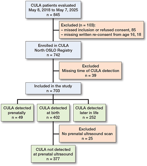
During the study period, we included 845 consecutive patients in the quality-assurance component of the CULA North Oslo Registry (Figure 1). Caregivers provided consent and completed the background questionnaire for the research component of the registry for 760 (90%) patients. Of these, we included 703 (93%) in our analysis as we excluded 18 patients for whom re-consent at 16 years had not been obtained and 39 patients with missing data regarding time of diagnosis. In 49 (7.0%) patients, the CULA was detected prenatally, while it was detected at birth in 402 (57%), and later in life in 252 (36%). 615 referrals (87%) originated from the South-Eastern Regional Health Authority. The other 3 Regional Health Authorities contributed 8, 33, and 47 referrals, which did not reflect the regions’ population sizes. Most patients with a CULA detected prenatally or at birth were evaluated in our hospital within the first months of life. Patients with a CULA detected later in life generally had less severe anomalies (Table 1).
Outcomes
The overall prenatal detection rate of CULAs visible at birth was 49/426 (12%; Figure 2, Table 2). 13 OMT phenotypes were detected prenatally, with detection rates ranging from 4.0% to 33%. Among phenotypes with large enough sample sizes for meaningful interpretation, we found the highest detection rates for congenital contractures (amyoplasia and distal arthrogryposis), reduction deficiencies (transverse, radial, and ulnar), and ulnar polydactyly (Figure 2, Table 2). 12 of 13 patients with prenatally detected symbrachydactyly lacked the whole hand or at least 3 fingers. All 8 patients with prenatally detected radial longitudinal deficiency lacked the whole radius and/or the thumb. Of the 14 patients with ulnar polydactyly, 11 had a floating, pedunculated 6th finger, while 3 had a more fully formed extra digit. According to the caregivers, the prenatal CULA diagnosis did not differ from the postnatal diagnosis in 36 of 49 patients.
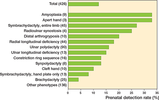
Figure 2. Congenital upper limb anomaly prenatal detection rates, overall and by phenotype. The total numbers of observed phenotypes are given in parentheses for each of the 13 phenotypes detected at prenatal ultrasound scans in the study population.
Among the 426 patients with a CULA visible at birth, 402 were born in 35 different Norwegian obstetric units, and 24 were born abroad. Prenatal detection was associated with birth in a university hospital, bilateral CULA, and musculoskeletal as well as internal organ anomalies, and VACTERL (vertebral anomalies, anal atresia, cardiac defects, tracheoesophageal fistula, renal anomalies, limb anomalies) association, but not with a plurality or a positive family history of congenital anomalies (Table 3). Adjustment for maternity hospital produced little change in odds ratios, with wider confidence intervals due to missing birthplace data for 28 patients (Table 3).
| Item | Detected prenatally (n = 49) |
Detected at birth (n = 377) |
Crude OR (CI) |
Adjusted OR a (CI) |
|||
| Caregiver-reported variables | |||||||
| Birth in a university hospital b | 33 (73) | 167 (47) | 3.1 (1.5–6.1) | ||||
| Twin or triplet c | 5 (10) | 18 (5) | 2.3 (0.80–6.4) | 2.2 (0.76–6.4) | |||
| Positive family history of | |||||||
| congenital anomalies d | 22 (46) | 125 (34) | 1.7 (0.92–3.1) | 1.8 (0.93–3.3) | |||
| CULA d | 14 (29) | 88 (24) | 1.3 (0.69–2.6) | 1.3 (0.66–2.7) | |||
| Assessor-reported variables | |||||||
| Bilateral CULA | 30 (61) | 169 (45) | 1.9 (1.1–3.6) | 2.1 (1.1–4.0) | |||
| Associated musculoskeletal anomalies | 18 (37) | 94 (25) | 1.8 (0.94–3.3) | 2.1 (1.1–4.0) | |||
| Lower limb | 12 (24) | 72 (19) | 1.4 (0.68–2.8) | 1.5 (0.76–3.2) | |||
| Spine | 3 (6) | 8 (2) | 3.0 (0.77–11) | 2.8 (0.69–11) | |||
| Associated other anomalies e | 21 (43) | 86 (23) | 2.5 (1.4–4.7) | 2.7 (1.4–5.1) | |||
| Heart | 9 (18) | 27 (7) | 2.9 (1.3–6.6) | 2.7 (1.2–6.3) | |||
| Syndrome | 10 (20) | 45 (12) | 1.9 (0.88–4.1) | 2.0 (0.90–4.3) | |||
| VACTERL association | 4 (8.2) | 7 (1.9) | 4.7 (1.3–17) | 4.5 (1.2–16) | |||
| a Adjusted for hospital type. b Missing data (n = 28): born abroad (n = 21), out-of-hospital births (n = 7). c Missing data (n = 1). d Missing data (n = 5). e All other anomalies besides the musculoskeletal system, including developmental delay. CI = 95% confidence interval, OR = odds ratio, VACTERL = vertebral anomalies, anal atresia, cardiac defects, tracheoesophageal fistula, renal anomalies, limb anomalies. |
|||||||
Discussion
Our study of 703 consecutive CULA patients provides new information on the time of diagnosis in a consecutive series of live-born children with CULA in a Western, sparsely populated country with a national prenatal screening program. We found that most patients had their CULA detected at birth, but a substantial proportion of patients were diagnosed much later in life. The overall prenatal detection rate was 12%, with the highest rates for congenital contractures, reduction deficiencies, and ulnar polydactyly. Patients with prenatally detected CULAs were more often born in a university hospital and presented more frequently with multiple congenital anomalies.
Not surprisingly, the prenatal CULA detection rate in our study population (12%) is lower than those reported from hand units in Australia and the USA for children with CULA. We assume this is mainly explained by the broad inclusion of all CULA phenotypes in our study in contrast to previous studies, including minor anomalies that are readily detected at birth but not during ultrasound scans, such as simple syndactyly and less severe reduction deficiencies (e.g., hypoplastic fingers including slight affection of forearm/wrist/carpus). An additional explanation might be the low population density and a decentralized healthcare system in Norway, in contrast to the high-volume, high-complexity hospitals where previous studies have been conducted. A registry-based Australian study reported an overall detection rate of 21%, but the registry included data on prenatal CULA detection for only 40% of the patients [5]. A study from the USA reported a detection rate of 31%, but it excluded minor CULAs that are difficult to detect on ultrasound [4]. The highest detection rate (42%) was reported in a study from the USA at a highly specialized tertiary referral center, where obstetrics and gynecology consultants performed all ultrasound examinations, and maternal-fetal medical specialists interpreted them [6]. It cannot be expected that less experienced assessors working in small obstetric units in rural areas in Norway obtain similar detection rates. However, the actual detection rate of CULA in the Norwegian national screening program is probably higher than 12%. Termination of pregnancies due to fetal anomaly (TOPFA) is not uncommon in Norway when multiple fetal anomalies are detected and has also been reported in cases of isolated limb reduction defects [15].
In our study, 73% of the parents confirmed the accuracy of the prenatal CULA diagnosis, which is comparable to the 58% reported in a USA study [4]. Similar to our findings, previous studies have indicated that amyoplasia, reduction deficiencies, and polydactyly have the highest prenatal detection rates [4-6], and that ulnar polydactyly is more often detected than radial polydactyly [4,5]. Despite this, it surprised us that ultrasounds failed to detect all 66 radial polydactylies and most reduction deformities.
Our finding that more CULAs were detected prenatally among patients born in university hospitals aligns with previous studies [3,6]. We could not assess associations between detection rates and Regional Health Authorities, as the number of referrals from the three Regional Health Authorities besides the South-Eastern reflected the local CULA surgical expertise more than the region’s population. Twin and triplet pregnancies involve more frequent ultrasound examinations, yet in our study, they were not associated with a higher prenatal CULA detection rate. This may reflect the small number of non-singletons in our study population, but it could also indicate the greater difficulty of visualizing all limb structures in multiple pregnancies. We did not find a higher prenatal detection rate in patients with a positive family history of CULA, despite its high prevalence in the study population. This may reflect that a large proportion of inherited CULA phenotypes visible at birth are minor finger defects that are not easily detected on prenatal ultrasound [16].
We found a higher prenatal detection rate in patients with bilateral CULAs than in those with unilateral CULAs. Many severe unilateral reduction deficiencies were not detected, possibly because the normal hand was described twice. Our study confirms previous findings of a higher detection rate in patients with associated anomalies, which, in turn, leads to a more meticulous upper limb examination [3,4,6,17]. Both findings are consistent with previous reports of a doubled likelihood of bilateral radial longitudinal deficiencies when part of a syndrome [17], and higher perinatal mortality in patients with bilateral limb anomalies [18,19]. VACTERL association had the strongest association with prenatal detection, which was not surprising as these patients often have multiple major anomalies of the internal organs.
In our study, only a small proportion of parents became aware of the CULA before birth, even for proximal deficiencies. A recent UK survey demonstrated considerable variation in parental preferences regarding whether a CULA diagnosis should be made prenatally or postnatally [20]. Regardless of timing, parents often fear associated undiagnosed anomalies, blame themselves, and worry about their child’s future physical and social functioning [20]. These concerns underscore the importance of congenital upper limb services in providing clear information regarding known associated anomalies, underlying conditions, and the generally high psychosocial functioning seen in children with CULA [21,22]. Our findings on time of diagnosis and prenatal detection rates provide clinicians with important, evidence-based expectations to communicate to families.
Strengths
The strengths of our study included comprehensive and structured data collection and CULA classification by senior consultant congenital hand surgeons over 7 years, with few missing data. The coverage of the research registry was high, and selection bias was minimal, as the registry included patients with all CULA diagnoses except tumorous dysplasias, ensuring high internal validity of the study. The external validity (for areas with similar demographics and health systems) was also high, as 87% of the patients in this study were consecutive admissions from a defined geographical region.
Limitations
The main limitation of our study was the unavailability of retrieving ultrasound reports from maternal medical records for verification of the caregiver-reported information concerning prenatal CULA diagnoses. Therefore, our study may suffer from parental misinterpretation bias or parental recall bias. Considering the median patient age of only a few months at registry enrollment, we assumed that caregiver‑reported information on whether a CULA was diagnosed at an ultrasound scan or not was sufficiently reliable for this study’s purpose, given the emotional impact typically associated with a prenatal anomaly diagnosis. On the other hand, parental assessments of the accuracy of the prenatal diagnosis may be less reliable. Obtaining ultrasonographic reports would have required additional written consent at the time of registry enrollment, and this procedure was not incorporated into the registry’s initial design. We considered retrospective collection of this data infeasible, as requiring new written consent from all 426 mothers would likely have caused response bias compromising the study’s sample size. Another study limitation was the relatively small study population, reflecting a sparsely populated country. Several rare phenotypes’ detection ranges had wide confidence intervals due to small sample sizes and must be interpreted with caution. Estimates for overall detection rate and for common phenotypes were considered acceptably precise.
Conclusion
7.0% of the upper limb anomalies were detected prenatally, 57% at birth, and 36% later in life. Prenatal detection rates were lower than those reported from larger non-European hand units, likely due to the inclusion of all upper limb anomaly diagnoses in this study and Norway’s decentralized prenatal care system.
- Ekblom A G, Laurell T, Arner M. Epidemiology of congenital upper limb anomalies in Stockholm, Sweden, 1997 to 2007: application of the Oberg, Manske, and Tonkin classification. J Hand Surg Am 2014; 39(2): 237-48. doi: 10.1016/j.jhsa.2013.11.014.
- Bae D S, Barnewolt C E, Jennings R W. Prenatal diagnosis and treatment of congenital differences of the hand and upper limb. J Bone Joint Surg Am 2009; 91(Suppl 4): 31-9. doi: 10.2106/jbjs.I.00072.
- Dicke J M, Piper S L, Goldfarb C A. The utility of ultrasound for the detection of fetal limb abnormalities: a 20-year single-center experience. Prenat Diagn 2015; 35(4): 348-53. doi: 10.1002/pd.4546.
- Gray B L, Calfee R P, Dicke J M, Steffen J, Goldfarb C A. The utility of prenatal ultrasound as a screening tool for upper extremity congenital anomalies. J Hand Surg Am 2013; 38(11): 2106-11. doi: 10.1016/j.jhsa.2013.08.091.
- O’Keefe D, Kennedy J, McCombe D, Coombs C, Hui L, Wilks D, et al. Pre-natal and post-natal diagnosis of congenital upper limb differences: the first 3 years of the Australian Hand Difference Register. J Paediatr Child Health 2022; 58(1): 122-8. doi: 10.1111/jpc.15673.
- Piper S L, Dicke J M, Wall L B, Shen T S, Goldfarb C A. Prenatal detection of upper limb differences with obstetric ultrasound. J Hand Surg Am 2015; 40(7): 1310-7.e3. doi: 10.1016/j.jhsa.2015.04.013.
- Vandenbroucke J P, von Elm E, Altman D G, Gøtzsche P C, Mulrow C D, Pocock S J, et al. Strengthening the Reporting of Observational Studies in Epidemiology (STROBE): explanation and elaboration. Ann Intern Med 2007; 147(8): W163-94. doi: 10.7326/0003-4819-147-8-200710160-00010-w1.
- Oslo University Hospital [Internet], Oslo [cited 2026 Jul 24]. Available from: https://www.oslo-universitetssykehus.no/avdelinger/ortopedisk-klinikk/ortopedisk-avdeling-rikshospitalet/nasjonal-behandlingstjeneste-for-dysmeli-i-overekstremiteten/
- McCombe D, Wall L, Goldfarb C, Hülsemann W, Sletten I N, Wilks D, et al. Congenital upper limb difference patient registries: characteristics, comparisons and recommendations. J Hand Surg Eur Vol 2025: 51(1): 111-18. doi: 10.1177/17531934251348360.
- Goldfarb C A, Ezaki M, Wall L B, Lam W L, Oberg K C. The Oberg–Manske-–Tonkin (OMT) classification of congenital upper extremities: update for 2020. J Hand Surg Am 2020; 45(6): 542-7. doi: 10.1016/j.jhsa.2020.01.002.
- Oberg K C. Classification of congenital upper limb anomalies: towards improved communication, diagnosis, and discovery. J Hand Surg Eur Vol 2019; 44(1): 4-14. doi: 10.1177/1753193418801280.
- Norwegian Directorate of Health [Internet], Oslo [cited 2026 Jul 24]. Available from: https://www.helsedirektoratet.no/retningslinjer/svangerskapsomsorgen/konsultasjoner-i-svangerskapsomsorgen#gravide-med-et-normalt-svangerskap-bor-fa-tilbud-om-et-basisprogram-praktisk-informasjon
- Blix E. Fødeinstitusjoner i Norge i Store medisinske leksikon [Internet], Oslo [cited 2026 July 24]. Available from: https://sml.snl.no/f%C3%B8deinstitusjoner_i_Norge
- Norwegian University of Science and Technology [Internet], Trondheim [cited 2026 July 24]. Available from: https://www.ntnu.no/videre/ultralydutdanning
- Klungsøyr K, Nordtveit T I, Kaastad T S, Solberg S, Sletten I N, Vik A K. Epidemiology of limb reduction defects as registered in the Medical Birth Registry of Norway, 1970–-2016: Population based study. PLoS One 2019; 14(7): e0219930. doi: 10.1371/journal.pone.0219930.
- Arduç A, van Dijk S J B, Ten Cate F J, van Doesburg M H M, Linskens I H, van Leeuwen E, et al. Phenotype-to-genotype description of prenatal suspected and postnatal discovered upper limb anomalies: a retrospective cohort study. Prenat Diagn 2025; 45(1): 3-14. doi: 10.1002/pd.6714.
- Forman M, Canizares M F, Bohn D, James M A, Samora J, Steinman S, et al. Association of radial longitudinal deficiency and thumb hypoplasia: an update using the CoULD Registry. J Bone Joint Surg Am 2020; 102(20): 1815-22. doi: 10.2106/jbjs.20.00281.
- Farr A, Wachutka E, Bettelheim D, Windsperger K, Farr S. Perinatal outcomes of infants with congenital limb malformations: an observational study from a tertiary referral center in Central Europe. BMC Pregnancy Childbirth 2020; 20(1): 35. doi: 10.1186/s12884-020-2720-x.
- Koskimies E, Lindfors N, Gissler M, Peltonen J, Nietosvaara Y. Congenital upper limb deficiencies and associated malformations in Finland: a population-based study. J Hand Surg Am 2011; 36(6): 1058-65. doi: 10.1016/j.jhsa.2011.03.015.
- Clelland A D, Lester R, Duncan Ó, Lam W L. Parental experience after diagnosis of a congenital upper limb difference: a national survey. J Hand Surg Eur Vol 2024; 49(11): 1327-33. doi: 10.1177/17531934241249014.
- Wilks D J, Cevik J, Kennedy J, Coombs C, Penington A, McCombe D. An analysis of the functional and psychosocial impact of congenital upper limb differences using PROMIS®: data from the Australian Hand Difference Register. J Hand Surg Eur Vol 2026; 51(1): 79-86. doi: 10.1177/17531934251355084.
- Bae D S, Canizares M F, Miller P E, Waters P M, Goldfarb C A. Functional impact of congenital hand differences: early results from the Congenital Upper Limb Differences (CoULD) Registry. J Hand Surg Am 2018; 43(4): 321-30. doi: 10.1016/j.jhsa.2017.10.006.