The Swedish experience of capital realignment surgery for slipped capital femoral epiphysis: results from a nationwide retrospective cohort of 28 patients
Jens NILSSON 1,2, Mikael LINDELL 3,4, Jakob ÖRTEGREN 1,2, Bengt HERNGREN 3,4, Margaretha STENMARKER 4–
6, Piotr MICHNO 3,4, and Carl Johan TIDERIUS 1,21 Department of Orthopedics, Skåne University Hospital, Lund; 2 Department of Clinical Sciences, Lund University, Lund; 3 Department of Orthopedics, Ryhov County Hospital, Jönköping; 4 Department of Biomedical and Clinical Sciences, Linköping University, Linköping; 5 Department of Pediatrics, Region Jönköping County, Jönköping; 6 Department of Pediatrics, Institute of Clinical Sciences, Sahlgrenska Academy, University of Gothenburg, Gothenburg, Sweden
ORCIDs, if available, can be found on the article page (https://www.actaorthop.org/actao/)
Background and purpose — Optimal treatment for slipped capital femoral epiphysis (SCFE) has been controversial for decades. Capital realignment surgery (predominant technique: modified Dunn) is a technique to restore anatomy, but it is associated with varying risks of avascular necrosis (AVN) of the femoral head. The aim of our study was to describe outcomes following capital realignment for severe SCFE in a nationwide cohort.
Methods — A longitudinal, nationwide, retrospective cohort study of SCFE patients treated with capital realignment between 2009 and 2021 was conducted, with radiographic and clinical follow-up 2–15 years after surgery. Primary radiographic outcome was AVN. Clinical outcomes were assessed using patient-reported outcome measures (the Copenhagen Hip and Groin Outcome Score [HAGOS] and the 12-item International Hip Outcome Tool [iHOT12]).
Results — 28 patients were included but 1 patient declined radiographic follow-up. 6/27 (22%, 95% confidence interval [CI] 11–41), developed AVN: 2/19 patients with stable SCFE and 4/8 patients with unstable SCFE. None of the 8 cases in which epiphyseal perfusion was monitored intraoperatively developed AVN. The median total HAGOS and iHOT12 scores were 77 (CI 56–94) and 76 (CI 72–88), respectively. When capital realignment was performed as primary treatment (< 1 month from diagnosis), the corresponding scores were 91 (CI 70–98) and 88 (CI 73–98).
Conclusion — The overall AVN rate was 22%. Our findings suggest that capital realignment should be reserved for stable SCFE and performed as primary treatment, with intraoperative epiphyseal perfusion monitoring.
Citation: Acta Orthopaedica 2026; 97: 503–509. DOI: https://doi.org/10.2340/17453674.2026.46429.
Copyright: © 2026 The Author(s). Published by MJS Publishing – Medical Journals Sweden, on behalf of the Nordic Orthopedic Federation. This is an Open Access article distributed under the terms of the Creative Commons Attribution 4.0 International License (https://creativecommons.org/licenses/by/4.0/)
Submitted: 2026-02-10. Accepted: 2026-06-26. Published: 2026-08-05.
Correspondence: jens.nilsson.8301@med.lu.se
Study design: JN, ML, BH, CJT. Data collection: JN, ML, BH, CJT. Review of radiographs: JN, ML. Data analysis: JN. Writing—original draft manuscript: JN. Writing—review and editing of manuscript: JN, ML, JÖ, BH, MS, PM, CJT.
The authors thank Olof Risto (OR), associate professor and orthopedic surgeon, who, together with the senior author (CJT), introduced capital realignment surgery as a treatment option for SCFE in Sweden.
They also thank Aleksandra Turkiewicz (AT), associate professor and statistician at Lund University, for statistical support.
Handling co-editors: Ilkka Helenius and Robin Christensen
Acta thanks Randall Loder, Jan Duedal Rölfing and other anonymous reviewers for help with peer review of this manuscript.
Optimal treatment for slipped capital femoral epiphysis (SCFE) has been controversial for decades. In-situ fixation is a percutaneous surgical procedure with negligible risk of avascular necrosis (AVN) of the femoral head [1]. However, in-situ fixation does not address the deformity which leads to worse long-term outcomes in severe slips [2]. Capital realignment is a technically demanding surgical procedure that aims to restore anatomy through open reduction of the epiphysis, thereby improving outcomes. Modified Dunn is the predominant technique. The procedure is associated with a risk of iatrogenic damage to the blood supply to the femoral head with subsequent AVN. Rates of AVN in the literature have ranged from negligible [3-5] to 50% or more [6,7].
Some authors argue for modified Dunn in all cases except mild SCFE, an approach that includes the majority of patients [5]. At the same time, in most pediatric orthopedic units, including Scandinavian ones, in-situ fixation is still predominant.
According to current Swedish guidelines [8], capital realignment is considered when the slip angle exceeds 50°. Since such cases are relatively few, the clinical experience with the procedure is still limited. The aim of our study was to describe outcomes in a longitudinal nationwide cohort to provide data for future decision-making regarding the management of severe SCFE. Specifically, we sought to study whether the risk of AVN can be justified by favorable patient-reported outcomes following restoration of proximal femoral anatomy.
Methods
Study design
We conducted a retrospective cohort study with active follow-up. We included all cases of SCFE treated with capital realignment in Sweden from the first case in 2009 to June 2021. The study is reported according to the STROBE guidelines.
Patients and data collection
Since 2007, children diagnosed with SCFE have been registered in a national Swedish Pediatric Orthopedic Quality register (SPOQ) [9]. There are approximately 50 cases annually and 4 in 5 have a mild or moderate slip (< 50°). The majority are treated with in-situ fixation [10].
Capital realignment as treatment for SCFE was independently introduced in Sweden by 2 consultant pediatric orthopedic surgeons from different orthopedic units (Malmö General Hospital, then Skåne University Hospital Lund, and Linköping University Hospital, then Ryhov County Hospital Jönköping). The first cases were performed in 2009. The following cases were performed at the above-mentioned surgeons’ units or by them at another unit. As there were no established national guidelines, it was at the discretion of the treating surgeon to determine whether capital realignment was indicated. National guidelines were first implemented in 2021.
All cases of capital realignment performed through June 2021 were identified by the treating surgeons. Patients were asked about study participation, including active follow-up with plain radiographs and patient-reported outcome measures (PROMs) if the hip was native at follow-up (defined as no total hip arthroplasty, THA). Study follow-up began in June 2023, and the minimum follow-up was 2 years—a period during which a possible AVN was expected to have manifested.
Data, including patients’ baseline characteristics and surgical reports, was retrospectively collected from medical records. The data was also checked against the national quality register (SPOQ). Previous radiographs (at diagnosis and after capital realignment) were reviewed. Slip angles were measured by 2 pediatric orthopedic surgeons with high intra- and interobserver agreement, as demonstrated in a previous publication [11]. On a frog leg lateral view (Lauenstein), the slip angle was measured according to the calcar femorale (CF) method [12]. Compared with the similar and internationally more commonly used lateral Southwick head–shaft angle (HSA) [13], accurate measurement can also be done in radiographs where only a small part of the proximal femur is visualized [11]. In cases with an additional Billing lateral view, the slip angle was also measured according to the Billing method [14]. Slip angles between ≥ 30° and ≤ 50° on lateral views (Lauenstein, Billing) were classified as moderate, and angles > 50° as severe [15,16]. If no lateral view was obtained at diagnosis (in cases with unstable slips, the child’s pain sometimes only allows for an AP image), the Southwick HSA on the anteroposterior (AP) view was used. HSA ≤ 110° on AP view was classified as severe [10].
Surgical technique
Capital realignment was performed both as the primary treatment for SCFE and at a later stage after percutaneous fixation at diagnosis. The predominant technique in our study was the modified Dunn procedure, which is based on the technique that was introduced by Dunn in 1964 [17]. The modified Dunn technique has been described in detail by Leunig et al. and Tannast et al. [3,5].
The hip was exposed via surgical dislocation. A retinacular soft tissue flap (posteriorly containing the major blood supply to the femoral head) was created by subperiosteal dissection around the femoral neck and the trochanteric region. The epiphysis was then mobilized through the physeal plane until it became mobile but still attached to the soft tissue flap. Callus formation was resected from the deformed metaphyseal stump, which was reshaped (including a slight shortening osteotomy) before the epiphysis was reduced and fixed. Alternatively, instead of prior mobilization through the physeal plane, some cases were operated on with a subcapital closing wedge osteotomy. As both techniques involve subcapital correction after the critical subperiosteal dissection, we assumed comparable risks of AVN and therefore included both techniques in the study.
The epiphyseal perfusion was monitored repeatedly throughout the capital realignment procedure. This was done either by drilling a small hole in the femoral head to observe for bleeding, or by placing an intracranial pressure (ICP) catheter in the femoral head to monitor pulsatile blood flow.
Outcomes
Follow-up radiographs were obtained at the nearest hospital (patients were distributed throughout the country) and then transferred to Skåne University Hospital. Radiographs were reviewed for AVN (primary outcome) and osteoarthritis (OA, secondary outcome), with OA defined as Kellgren–Lawrence grade ≥ 2 [18,19]. In patients who had undergone hip replacement by follow-up, the indication for conversion to THA was recorded (AVN or OA). The α-angle, a measure of cam morphology (secondary outcome), was measured on AP radiographs. As suggested in a cross-sectional study on a large Scandinavian cohort by Gosvig et al., a pathological cutoff was set at a greater angle for males (83°) than for females (57°) due to the inherent anatomical difference between the sexes [20].
Clinical outcomes were assessed with PROMs. Questionnaires were sent to the patients by letter. The Copenhagen Hip and Groin Outcome Score (HAGOS) and the 12-item International Hip Outcome Tool (iHOT12) were used in Swedish versions that have been validated in patients 15 to 75 years old [21,22]. Both HAGOS and iHOT12 have been used to assess clinical outcomes after SCFE in previous publications [23,24]. HAGOS scores were calculated for each subscale and as a total score, and the iHOT12 score was calculated as the mean of the 12 items (ranging from 0 = worst possible to 100 = best possible).
Statistics
Continuous descriptive data was summarized as medians with interquartile ranges (IQR). All estimates were accompanied by 95% confidence intervals (CIs). CIs for binary outcomes (AVN, OA, pathological AP α-angle; yes/no) were calculated using the Wilson score method. CIs for continuous outcomes summarized by medians (AP α-angle, PROM scores) were obtained by bootstrap resampling (5,000 resamples). Correlations between continuous outcomes were analyzed using Spearman’s correlation coefficient (rs). In exploratory subgroup analyses, primary outcomes were compared between groups defined by variables hypothesized to influence the outcome, such as clinical classification of the slip (stable/unstable, the latter meaning patients unable to ambulate even with crutches) [25]. Between-group differences in AVN rate were estimated as risk differences (percentage points), with CIs calculated using the Newcombe method. For PROM scores, between-group differences were estimated as the Hodges–Lehmann location shift. IBM SPSS Statistics version 29 (IBM Corp, Armonk, NY, USA) was used for the statistical analyses.
Ethics, data sharing, and funding
Ethical approval was obtained from the Swedish Ethical Review Authority with diary number 2021-00056 (March 3, 2021). All potential study participants who were reachable were informed by letter. Written informed consents were obtained.
All documents with original data of relevance will be stored for a minimum of 10 years at the Department of Orthopedics, Skåne University Hospital, Lund, Sweden. Pseudonymized study data can be obtained upon request.
Research grants were received from Erik och Angelica Sparres forskningsstiftelse (Lund, Sweden), Kockska stiftelserna (Trelleborg, Sweden), and Futurum – akademin för hälsa och vård (Jönköping County Council, Sweden). Complete disclosure of interest forms according to ICMJE are available on the article page, doi: 10.2340/17453674.2026.46429
Results
28 patients were included in the study (Figure 1). The patients were evaluated 2–15 years after the capital realignment surgery. Study follow-up was complete for 27 patients (1 patient declined to undergo new radiographs). Descriptive characteristics of the cohort are given in Table 1.
| Factors | n | Cohort distribution | ||
| Diagnosis-related characteristics | ||||
| Sex (male/female) | 28 | 16/12 | ||
| Affected side (left/right) | 28 | 19/9 | ||
| Age (years) | 28 | 13 (9, 11–14, 17) | ||
| Body mass index | 20 | 24 (16, 19–26, 31) | ||
| Clinical classification (stable/unstable) | 28 | 20/8 | ||
| Severity of slip (moderate/severe a) | 28 | 4/24 | ||
| Slip angle frog leg lateral, CF (°) | 24 | 61 (46, 53–74, 89) | ||
| Realignment-related characteristics | ||||
| Age at capital realignment (years) | 28 | 13 (9, 12–15, 21) | ||
| Treating orthopedic unit (Lund-Malmö/Linköping-Jönköping) | 28 | 9/19 | ||
| Primary capital realignment (yes b/no c) | 28 | 17/11 | ||
| Capital realignment technique (modified Dunn/closing wedge) | 28 | 20/8 | ||
| Reduction of the CF slip angle if comparable frog leg lateral pre- and postoperatively (°) | 16 | 53 (40, 45–65, 72) | ||
| Use of ICP device to intraoperatively monitor epiphyseal perfusion (yes/no) | 28 | 8/20 | ||
| Perfusion on postoperative bone scintigraphy if performed (normal/reduced) | 22 | 15/7 | ||
| Time from capital realignment to radiographs at follow-up if native hip (months) | 20 | 106 (24, 57–151, 181) | ||
| Time from capital realignment to PROMs at follow-up if native hip (months) | 21 | 111 (24, 60–147, 170) | ||
| a Slip angles > 50° according to CF and/or Billing. AP Southwick HSA of ≤ 110° in cases with no lateral view radiographs at diagnosis. 1 slip was assessed as severe based on MRI at diagnosis as no radiographs were obtained at that time. b 1–27 days from diagnosis. c 4–76 months from diagnosis/primary treatment. CF = calcar femorale , ICP = intracranial pressure, IQR = interquartile range, PROMs = patient-reported outcome measures. |
||||
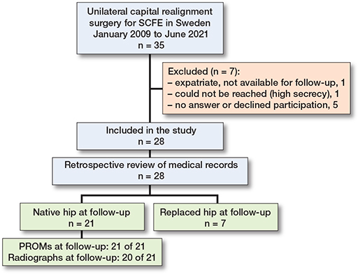
Figure 1. Flowchart of included patients. PROMs = patient-reported outcome measures, SCFE = slipped capital femoral epiphysis.
Outcomes
Of 27 patients, 6 (22%, CI 11–41) developed AVN. Of the patients with AVN, 5 had undergone THA by follow-up (Table 2). Figure 2 demonstrates such a case, while Figure 3 exemplifies a case with an excellent outcome following modified Dunn. OA developed in 4 of 27 patients (15%, CI 6–33). In 2 of these patients, OA led to conversion to THA.
| Item | n (n a) | Outcome (CI) | ||
| Radiographic outcomes (N = 27) | ||||
| Avascular necrosis | 6 (5) | 22% (11–41) | ||
| Osteoarthritis | 4 (2) | 15% (6–33) | ||
| PROMs (native hips) | ||||
| HAGOS total score, n median | 21 | 77 (56–94) | ||
| iHOT12 score, n median | 21 | 76 (72–88) | ||
| a number of cases converted to total hip arthroplasty in parentheses. CI = 95% confidence interval, HAGOS = Copenhagen Hip and Groin Outcome Score, iHOT12 = 12-item International Hip Outcome Tool, N = total number of patients with available data (1 patient with native hip at study follow-up declined new radiographs), PROMs = patient-reported outcome measures. |
||||
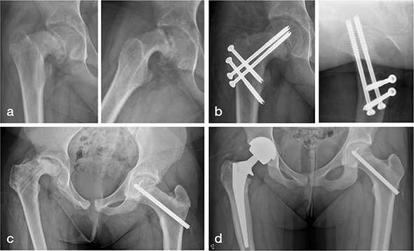
Figure 2. Radiographs (a) of a 12-year-old girl with a 7-month history of hip-related symptoms show a severe, stable right-sided SCFE (slip angle 72°). The girl underwent capital realignment using the modified Dunn technique as primary treatment (b). The patient developed avascular necrosis, and the screws were removed (c). The hip was converted to a THA 16 months after the modified Dunn (d).
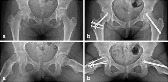
Figure 3. Radiographs (a) of a 12-year-old girl with a 4-month history of hip-related symptoms show a right-sided, severe, stable SCFE (slip angle 60°). The girl underwent modified Dunn surgery at diagnosis, with simultaneous prophylactic nailing on the contralateral side. At 2-year follow-up, the patient reported excellent outcome with normalized hip function (b).
Radiographs at follow-up showed a pathological AP α-angle in 12 of 19 patients (63%, CI 41–81; accurate measurement was not possible in 1 case due to AVN). The median AP α-angle was 89° (CI 49–100).
The median total HAGOS score at follow-up was 77 (CI 56–94), and the median iHOT12 score was 76 (CI 72–88) (see Table 2). PROM score distributions for each HAGOS subscale and the iHOT12 score showed considerable variation and are presented alongside reference scores from asymptomatic young populations [26,27] in Figure 4. The correlation between α-angle and PROM scores was rs –0.34 (CI –0.75 to 0.17) for the total HAGOS score and rs –0.38 (CI –0.74 to 0.10) for the iHOT12 score.
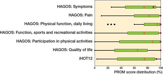
Figure 4. Patient-reported outcome score distributions in the cohort for each Copenhagen Hip and Groin Outcome Score (HAGOS) subscale and the 12-item International Hip Outcome Tool (iHOT12) score. Purple markers = reference scores from asymptomatic young populationsa. Vertical red line = median, box = upper and lower IQR, whisker = values within 1.5 x IQR, and black dots = outliers.
a Median HAGOS scores in asymptomatic soccer players (mean age 23.6) with no pain in the previous season [26]. Normative mean iHOT score in patients < 40 years with no history of chronic hip pain or previous hip surgery [27].
Exploratory analyses of differences in primary outcomes across subgroups are presented in Tables 3 and 4. 4 of 8 patients with unstable SCFE developed AVN. There were no cases of AVN among the 8 patients in whom the epiphyseal perfusion was monitored with an ICP device during surgery. When capital realignment was used as the primary treatment, the median total HAGOS and iHOT12 scores were 91 (CI 70–98) and 88 (CI 73–98), respectively.
| Subgroups | n of N a with AVN | Between-group risk difference in percentage points (CI) | ||
| Clinical classification | ||||
| Stable | 2 of 19 | 40 (4 to 69) | ||
| Unstable | 4 of 8 | |||
| Use of intraoperative ICP monitor | ||||
| Yes | 0 of 8 | 32 (–5 to 54) | ||
| No | 6 of 19 | |||
| Type of capital realignment | ||||
| Modified Dunn | 4 of 19 | 4 (–25 to 40) | ||
| Closing wedge | 2 of 8 | |||
| Primary capital realignment | ||||
| Yes (< 1 month from diagnosis) | 4 of 17 | 4 (–31 to 30) | ||
| No (> 4 months from diagnosis) | 2 of 10 | |||
| Orthopedic unit | ||||
| Lund/Malmö | 1 of 9 | 17 (–19 to 42) | ||
| Linköping/Jönköping | 5 of 18 | |||
| Learning curve per unit | ||||
| 1st half | 4 of 14 | 13 (–19 to 42) | ||
| 2nd half | 2 of 13 | |||
| a n of N: n = number of patients with avascular necrosis (AVN); N = total number of patients in the subgroup. CI = 95% confidence interval, ICP = intracranial pressure. |
||||
Discussion
To our knowledge, this is the first study of capital realignment surgery in Scandinavia, presenting Sweden’s initial 12-year experience with this debated procedure. The 11% AVN rate in stable SCFE, together with the favorable patient-reported outcome in cases operated on within 1 month from diagnosis, may support the use of capital realignment for severe slips. However, the threshold for what is considered an acceptable risk of AVN varies, and long-term comparative studies of in-situ fixation vs capital realignment are needed.
AVN rates following SCFE treated with percutaneous fixation in Sweden are low. In a nationwide cohort including all SCFE cases from 2007 to 2013, only 5 of 380 (1%) hips with a stable slip developed AVN, including 2 of 41 (5%) severe slips. For unstable slips, AVN developed in 11 of 56 (20%) hips [28]. The overall AVN rate after capital realignment in our cohort (22%) fell within the previously reported extremes of the modified Dunn procedure. It was higher than the overall rate of 14% reported in a previously published meta-analysis [29], mainly because of the unstable slips in our cohort. Regarding stable slips, our AVN rate matched that in the meta-analysis: 11%. In our study, 17 of 19 stable cases had no AVN at follow-up. Still, the correct number is probably 18 of 20 because the patient who declined follow-up radiographs had a native hip and reported a good outcome > 10 years after capital realignment.
Regarding unstable slips, some authors have recommended a modified Dunn procedure to reduce the risk of AVN [30], while others have warned of an increased risk [7]. AVN following unstable slips was frequent in our cohort: 4 of 8 cases. Consequently and according to current Swedish guidelines [8], we no longer treat unstable slips with modified Dunn as first surgical procedure. In stable SCFE, in-situ fixation is recommended for mild to moderate slips whereas capital realignment is considered only in severe cases.
There were no cases of AVN among the 8 cases in which an ICP catheter was used to monitor femoral head perfusion intraoperatively, compared with 6 of 19 cases in which no ICP catheter was used. The subgroup analyses were exploratory and should be interpreted accordingly. However, the use of an ICP device has also been supported previously; Birke retrospectively assessed 217 consecutive modified Dunn procedures and found that, with intraoperative monitoring (ICP device), the AVN rate after stable SCFE decreased from 21% to 0%. In unstable SCFE, the rate remained unchanged [31].
Reported long-term outcomes following in-situ fixation are significantly worse in severe slips compared with mild and moderate slips. At a mean follow-up of 18 years, de Poorter et al. found that 75% of the patients with severe slips had severe OA and their patient-reported outcome was poor [2]. The main goal of capital realignment is improved clinical outcomes. Different outcome scores appear in the literature, whereby comparison across studies is difficult. In a meta-analysis using the Harris Hip Score, the overall mean score was excellent [29], whereas our PROMs were fair to good. However, our PROM scores improved to very good when capital realignment was performed as the primary treatment. Comparable iHOT12 scores following the modified Dunn procedure have been previously reported at shorter follow-up (1 year) [32]. When capital realignment was performed at a later stage, the median PROM scores in our cohort were poor.
SCFE is associated with an increased incidence of femoroacetabular impingement due to cam deformity [33], and FAI is a risk factor for developing OA [34]. In our study, capital realignment did not eliminate the risk of cam morphology. There was a negative trend between a high α-angle and a worse clinical outcome. The weak correlation may reflect a Type II error, a short follow-up time, or both. It is also possible that factors other than the α-angle are more important for the clinical outcome. Delayed capital realignment was associated with OA (all 4 cases), which might be explained by the degenerative process having already started at the time of realignment surgery.
Strengths
The main strength of our study is its nationwide scope. Furthermore, it is a longitudinal study with few patients lost to follow-up.
Limitations
The study describes outcomes from the earliest to the later cases of a technically demanding procedure, thereby reflecting the surgeons’ learning curve. The study has selection bias, since the indication for capital realignment was at the discretion of the treating surgeon. A few cases involved capital realignment using a subcapital closing wedge osteotomy, which does not strictly adhere to the modified Dunn technique. The absence of a control group limits the ability to compare outcomes with alternative treatment options. The relatively small cohort size reduces the study’s statistical power, and the subgroup findings should be interpreted as exploratory. It is possible that a larger cohort would have provided more robust evidence on the use of an ICP device. The follow-up time may have been too short for some patients to evaluate the effect of FAI on both symptoms and OA onset.
Conclusion
The overall AVN rate was 22%, 11% in stable SCFE, and 50% in unstable SCFE. There were no cases of AVN when the epiphyseal perfusion was monitored with an ICP device during surgery. PROMs were fair to good but improved to very good when capital realignment was used as primary treatment.
In perspective, our findings suggest that capital realignment should be reserved for stable SCFE and used as primary treatment with intraoperative epiphyseal perfusion monitoring. However, further long-term comparative studies are warranted.
- Naseem H, Chatterji S, Tsang K, Hakimi M, Chytas A, Alshryda S. Treatment of stable slipped capital femoral epiphysis: systematic review and exploratory patient level analysis. J Orthop Traumatol 2017; 18(4): 379-94. doi: 10.1007/s10195-017-0469-4.
- de Poorter J J, Beunder T J, Gareb B, Oostenbroek H J, Bessems G H, van der Lugt J C, et al. Long-term outcomes of slipped capital femoral epiphysis treated with in situ pinning. J Child Orthop 2016; 10(5): 371-9. doi: 10.1007/s11832-016-0759-z.
- Leunig M, Slongo T, Kleinschmidt M, Ganz R. Subcapital correction osteotomy in slipped capital femoral epiphysis by means of surgical hip dislocation. Oper Orthop Traumatol 2007; 19(4): 389-410. doi: 10.1007/s00064-007-1213-7.
- Ziebarth K, Zilkens C, Spencer S, Leunig M, Ganz R, Kim Y-J. Capital realignment for moderate and severe SCFE using a modified Dunn procedure. Clin Orthop Rel Res 2009; 467(3): 704-16. doi: 10.1007/s11999-008-0687-4.
- Tannast M, Jost L M, Lerch T D, Schmaranzer F, Ziebarth K, Siebenrock K A. The modified Dunn procedure for slipped capital femoral epiphysis: the Bernese experience. J Child Orthop 2017; 11(2): 138-46. doi: 10.1302/1863-2548-11-170046.
- Masquijo J J, Allende V, D’Elia M, Miranda G, Fernández C A. Treatment of slipped capital femoral epiphysis with the modified Dunn procedure: a multicenter study. J Pediatr Orthop 2019; 39(2): 71-5. doi: 10.1097/bpo.0000000000000936.
- Alves C, Steele M, Narayanan U, Howard A, Alman B, Wright J G. Open reduction and internal fixation of unstable slipped capital femoral epiphysis by means of surgical dislocation does not decrease the rate of avascular necrosis: a preliminary study. J Child Orthop 2012; 6(4): 277-83. doi: 10.1007/s11832-012-0423-1.
- Beslutsstöd för höftfyseolys, SBOF Svensk BarnOrtopedisk Förening [in Swedish]. Available from: https://slf.se/sof/app/uploads/2021/02/sbof-spoq-beslutsstoxxd-hoxxftfyseolys-2021-02-26.pdf (revised version adopted 2021).
- SPOQ Svenskt Pediatriskt Ortopediskt Qvalitetsregister, Registercentrum Västra Götaland [in Swedish]. Available from: https://spoq.registercentrum.se/ (accessed 2021).
- Herngren B, Stenmarker M, Vavruch L, Hagglund G. Slipped capital femoral epiphysis: a population-based study. BMC Musculoskelet Disord 2017; 18: 1-12. doi: 10.1186/s12891-017-1665-3.
- Lindell M, Nilsson J, Herngren B, Ortegren J, Stenmarker M, Tiderius C J, et al. Comparison of different radiographic methods to measure the slip angle in children with slipped capital femoral epiphysis (SCFE). Acta Radiol 2024; 65(9): 1109-14. doi: 10.1177/02841851241271999.
- Hansson L I, Hägglund G, Ordeberg G, Sandström S. The calcar femorale as a landmark in hip physiolysis. Acta Orthop Scand 1988; 59(2): 134-8. doi: 10.3109/17453678809169693.
- Southwick W O. Osteotomy through the lesser trochanter for slipped capital femoral epiphysis. J Bone Joint Surg Am 1967; 49(5): 807-35.
- Billing L, Bogren H G, Wallin J. Reliable X-ray diagnosis of slipped capital femoral epiphysis by combining the conventional and a new simplified geometrical method. Pediatr Radiol 2002; 32(6): 423-30. doi: 10.1007/s00247-002-0697-4.
- Hansson G, Billing L, Högstedt B, Jerre R, Wallin J. Long-term results after nailing in situ of slipped upper femoral epiphysis: a 30-year follow-up of 59 hips. J Bone Joint Surg Br 1998; 80(1): 70-7. doi: 10.1302/0301-620x.80b1.7268.
- Loder R T, Starnes T, Dikos G, Aronsson D D. Demographic predictors of severity of stable slipped capital femoral epiphyses. J Bone Joint Surg Am 2006; 88(1): 97-105. doi: 10.2106/JBJS.E.00069.
- Dunn D M. The treatment of adolescent slipping of the upper femoral epiphysis. J Bone Joint Surg Br. 1964; 46(4): 621-9.
- Kellgren J H, Lawrence J S. Radiological assessment of osteo-arthrosis. Ann Rheum Dis 1957; 16(4): 494-502.
- Kohn M D, Sassoon A A, Fernando N D. Classifications in brief: Kellgren–Lawrence classification of osteoarthritis. Clin Orthop Relat Res 2016; 474(8): 1886-93. doi: 10.1007/s11999-016-4732-4.
- Gosvig K, Jacobsen S, Sonne-Holm S, Gebuhr P. The prevalence of cam-type deformity of the hip joint: a survey of 4151 subjects of the Copenhagen Osteoarthritis Study. Acta Radiol 2008; 49(4): 436-41. doi: 10.1080/02841850801935567.
- Thomeé R, Jónasson P, Thorborg K, Sansone M, Ahldén M, Thomeé C, et al. Cross-cultural adaptation to Swedish and validation of the Copenhagen Hip and Groin Outcome Score (HAGOS) for pain, symptoms and physical function in patients with hip and groin disability due to femoro-acetabular impingement. Knee Surg Sports Traumatol Arthrosc 2014; 22(4): 835-42. doi: 10.1007/s00167-013-2721-7.
- Jónasson P, Baranto A, Karlsson J, Swärd L, Sansone M, Thomeé C, et al. A standardised outcome measure of pain, symptoms and physical function in patients with hip and groin disability due to femoro-acetabular impingement: cross-cultural adaptation and validation of the international Hip Outcome Tool (iHOT12) in Swedish. Knee Surg Sports Traumatol Arthrosc 2014; 22(4): 826-34. doi: 10.1007/s00167-013-2710-x.
- Örtegren J, Peterson P, Svensson J, Tiderius C J. Persisting CAM deformity is associated with early cartilage degeneration after slipped capital femoral epiphysis: 11-year follow-up including dGEMRIC. Osteoarthritis Cartilage 2018; 26(4): 557-63. doi: 10.1016/j.joca.2018.01.013.
- Gelink A, Cúneo A, Silveri C, Tiderius C J, Loder R, von Heideken J. Valgus slipped capital femoral epiphysis: presentation, treatment, and clinical outcomes using patient-reported measurements. J Pediatr Orthop B 2021; 30(2): 111-15. doi: 10.1097/BPB.0000000000000736.
- Loder R T, Richards B S, Shapiro P S, Reznick L R, Aronson D D. Acute slipped capital femoral epiphysis: the importance of physeal stability. J Bone Joint Surg Am 1993; 75(8): 1134-40. doi: 10.2106/00004623-199308000-00002.
- Thorborg K, Branci S, Stensbirk F, Jensen J, Hölmich P. Copenhagen Hip and Groin Outcome Score (HAGOS) in male soccer: reference values for hip and groin injury-free players. Br J Sports Med 2014; 48(7): 557-9. doi: 10.1136/bjsports-2013-092607.
- Sharfman Z T, Safran N, Amar E, Varshneya K, Safran M R, Rath E. Age-adjusted normative values for hip patient-reported outcome measures. Am J Sports Med 2022; 50(1): 79-84. doi: 10.1177/03635465211056666.
- Herngren B, Stenmarker M, Enskär K, Hägglund G. Outcomes after slipped capital femoral epiphysis: a population-based study with three-year follow-up. J Child Orthop 2018; 12(5): 434-43. doi: 10.1302/1863-2548.12.180067.
- Jauregui J J, Shaw N M, Weir T B, Barvarz S A, McClure P K. Risk of avascular necrosis with the modified Dunn procedure in SCFE patients: a meta-analysis. Children (Basel) 2022; 9(11). doi: 10.3390/children9111680.
- Ziebarth K, Leunig M, Slongo T, Kim Y J, Ganz R. Slipped capital femoral epiphysis: relevant pathophysiological findings with open surgery. Clin Orthop Relat Res 2013; 471(7): 2156-62. doi: 10.1007/s11999-013-2818-9.
- Birke O, George J S, Gibbons P J, Little D G. The modified Dunn procedure can be performed safely in stable slipped capital femoral epiphysis but does not alter avascular necrosis rates in unstable cases: a large single-centre cohort study. J Child Orthop 2021; 15(5): 479-87. doi: 10.1302/1863-2548.15.210106.
- Elsiofy A, Elsherif M, Eladawy M F, Mahmoud T, Sakr A F. How safe and technical is modified Dunn osteotomy in the management of patients with SCFE: a clinical trial with short-term follow-up. Adv Orthop 2023; 2023:2742083. doi: 10.1155/2023/2742083.
- Terjesen T, Wensaas A. Prognostic factors for long-term outcome of chronic slipped capital femoral epiphysis treated with fixation in situ. J Child Orthop 2017; 11(2): 114-19. doi: 10.1302/1863-2548-11-160285.
- Sankar W N, Nevitt M, Parvizi J, Felson D T, Agricola R, Leunig M. Femoroacetabular impingement: defining the condition and its role in the pathophysiology of osteoarthritis. J Am Acad Orthop Surg 2013; 21(Suppl 1): S7-s15. doi: 10.5435/jaaos-21-07-s7.